Monographs: Radiopharmaceuticals: General monograph: 8. IDENTIFICATION
8.1 Measurement of radioactivity
The radioactivity of a radiopharmaceutical preparation is stated at a given date and, if necessary, time. The absolute measurement of the radioactivity of a given sample may be carried out if the decay scheme of the radionuclide is known but, in practice, many corrections are required to obtain accurate results. For this reason, it is common to carry out the measurement with the aid of a primary standard source. Primary standards may not be available for short-lived radionuclides, e.g. β+ emitters. Measuring instruments are calibrated using suitable standards for the particular radionuclides. Standards are available from the laboratories recognized by the competent authority. Ionization chambers and Geiger-Müller counters may be used to measure β- and β-/γ emitters; scintillation or semiconductor counters or ionization chambers may be used for measuring γ emitters; low-energy β- emitters require a liquid-scintillation counter. For the detection and measurement of α emitters, specialized equipment and techniques are required. For an accurate comparison of radioactive sources, it is essential for samples and standards to be measured under similar geometrical conditions.
Low-energy β- emitters may be measured by liquid-scintillation counting. The sample is dissolved in a solution containing one or often two organic fluorescent substances (primary and secondary scintillators) which convert part of the energy of disintegration into photons of light which are detected by a photomultiplier and converted into electrical impulses. When using a liquid-scintillation counter, comparative measurements are corrected for light-quenching effects. Direct measurements are made, wherever possible, under similar conditions, (e.g. volumes and type of solutions) for the source to be examined and for the standard source.
All measurements of radioactivity must be corrected by subtracting the background due to radioactivity in the environment and due to spurious signals generated in the equipment itself.
With some equipment, when measurements are made at high levels of radioactivity, it may be necessary to correct for the losses due to the finite resolving time of the detector and its associated electronic equipment (dead time). For a counting system with a fixed dead time τ following each count, the correction is:
N = Nobs/(1 – Nobs x τ)
N = the true count rate per second,
Nobs = the observed count rate per second,
τ = the dead time, in seconds.
With some equipment this correction is made automatically. Corrections for loss by dead time must be made before the correction for background radiation.
8.2 Correction for decay during measurement
If the time of an individual measurement, tm is not negligible short compared with the half-life of the radionuclide, T1/2, the decay during this measurement time must be taken into account. After having corrected the instrument reading (count rate, ionization current, etc.) for background and, if necessary, for losses due to electronic effects, the decay correction during measurement time is:
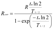
Rcorr = instrument reading corrected to the beginning of the individual measurement,
R = instrument reading before decay correction, but already corrected for background, etc.
8.3 Statistics of radioactivity measurement
The results of radioactivity determination show variation, which mainly are derived from the random nature of nuclear transformation. A sufficient number of counts must be registered in order to compensate for variations in the number of transformations per unit of time. The standard deviation is the square root of the counts, so at least 10 000 counts are necessary to obtain a relative standard deviation of not more than 1% (confidence interval: 1 sigma).
All statements of radioactive content should be accompanied by a statement of the date and, if necessary, the time at which the measurement was made. This statement of the radioactive content must be made with reference to a time zone (e.g., GMT, CET) or UTC with offset (e.g., UTC+02:00). The radioactivity at other times can be calculated from the exponential equation or from tables.
The radioactivity of a solution is expressed per unit volume to indicate the radioactive concentration.
8.4 Measurement of half-life (T1/2)
The T1/2 is measured with a suitable radiation detector (See Measurement of Radioactivity.)
The exponential decay (decay curve) is described by the equation:
At = Ao e-λt
At = the radioactivity at time t,
Ao = the radioactivity at time t = 0,
λ = the decay constant characteristic of each radionuclide,
e = the base of natural (Napierian) logarithm.
The half-life (T1/2) is related to the decay constant (λ) by the equation:
T1/2 = 0.693/ λ
A radionuclide is generally identified by its half-life or by the nature and energy of its radiation or radiations emitted or by both, as prescribed in the monograph.
The radiopharmaceutical preparation to be tested is used as such or diluted or dried in a capsule after appropriate dilution. The radioactivity chosen, having regard to experimental conditions, must be of a sufficiently high level to allow detection during several estimated T1/2, but not too high to minimize count rate losses, for example due to dead time.
The radioactive source is prepared in a manner that will avoid loss of material during handling. If it is a liquid (solution), it is contained in bottles or sealed tubes. If it is a solid (residue from drying in a capsule), it is protected by a cover consisting of a sheet of adhesive cellulose acetate or of some other material.
Perform the measurements using an appropriately calibrated instrument, such as a quantitative radioactivity detector, provided the quantity of radioactivity is within the linear range of the instrument throughout the measurements.
The same source is measured in the same geometry and at intervals usually corresponding to half of the estimated half-life throughout a time equal to about three half-lives. The performance of the apparatus is checked using a source of long T1/2 and, if necessary, corrections for any changes in the count rate have to be applied (see Measurement of Radioactivity).
A graph can be drawn with time as the abscissa and the logarithm of the relative instrument reading (e.g. count rate) as the ordinate. The calculated T1/2 should not differ by more than 5% from the expected T1/2, unless otherwise stated in the monograph.
8.5 Determination of the nature and energy of the radiation
The nature and energy of the radiation emitted may be determined by several procedures including the construction of an attenuation curve and the use of spectrometry. The attenuation curve can be used for analysis of β radiation. Spectrometry is mostly used for identification of γ rays and detectable X-rays.
The attenuation curve is drawn for pure electron emitters when no spectrometer for beta rays is available or for beta/gamma emitters when no spectrometer for gamma rays is available. This method of estimating the maximum energy of beta radiation gives only an approximate value. The source, suitably mounted to at fixed geometry, is placed in front of the thin window of a Geiger-Müller counter or a proportional counter. The source is protected as described above. The count rate of the source is then measured. Between the source and the counter are placed, in succession, at least six aluminum screens of increasing mass per unit area. Within such limits that with a pure beta emitter this count rate is not affected by the addition of further screens. The screens are inserted in such a manner that constant geometrical conditions are maintained. A graph is drawn showing the mass per unit area of the screen expressed in milligrams per square centimeter as the abscissa and, the logarithm of the count rate as the ordinate for each screen examined. A graph is drawn in the same manner for a standardized preparation. The mass attenuation coefficients are calculated from the median parts of the curves, which are practically rectilinear.
The mass attenuation coefficient µm, expressed in square centimeters per milligram, depends on the energy spectrum of the beta radiation and on the nature and the physical properties of the screen. It therefore allows beta emitters to be identified. It is calculated using the equation:
µm = (2.303(log A1 – logA2))/(m1 + m2)
m1 = mass per unit area of the lightest screen,
m2 = mass per unit area of the heaviest screen, m1 and m2 being within the rectilinear part of the curve,
A1 = count rate for mass per unit area m1,
A2 = count rate for mass per unit area m2.
The mass attenuation coefficient µm, thus calculated, does not differ by more than 10% from the coefficient obtained under identical conditions using a standardized preparation of the same radionuclide.
The range of β- particles is an additional parameter which can be used for the determination of the β- energy. It is obtained from the graph described above as the mass per unit area corresponding to the intersection of the extrapolations of the descending rectilinear part of the attenuation curve and the horizontal line of background radioactivity.
Liquid scintillation counting may be used to obtain spectra of α and β- emitters (see Measurement of Radioactivity).
Gamma spectrometry is used to identify radionuclides by the energy and intensity of their γ rays and X-rays.
The preferred detectors for γ and X-ray spectrometry are semiconductors such as germanium or cadmium-zinc-telluride (CZT). A thallium-activated sodium iodide scintillation detector is also used but this has a much lower energy resolution.
The gamma detector has to be calibrated using standard sources because the detection efficiency is a function of the energy of the γ and X-rays as well as the form of the source and the source-to-detector distance. The detection efficiency may be measured using a calibrated source of the radionuclide to be measured or, for more general work, a graph of efficiency against γ and X-ray energy may be constructed from a series of calibrated sources of various radionuclides.
The γ and X-ray spectrum of a radionuclide which emits γ and X-rays is unique to that nuclide (apart from positron emitting radionuclides) and is characterized by the energies and the number of photons of particular energies emitted per transformation from one energy level to another energy level. This property contributes to the identification of radionuclides present in a source and to their quantification. It allows the estimation of the degree of radionuclidic impurity by detecting peaks other than those expected. Positron emitting radionuclides can only be identified through their difference in half-life.
It is possible to establish the rate of the decay of radioactivity using γ-spectrometry since the peaks diminish in amplitude as a function of the T1/2. If, in such a source, a radioactive impurity with a different T1/2 is present, it is possible to detect the latter by identification of the characteristic peak or peaks whose amplitudes decrease at a different rate from that expected for the particular radionuclide. A determination of the T1/2 of the additional peaks by repeated measurements of the sample will help to identify the impurity.
The table of physical characteristics of radionuclides (Annex 3) summarizes the most commonly accepted physical characteristics of radionuclides used in radiopharmaceutical preparations. The table also states the physical characteristics of the main potential impurities of the radionuclides.
By "transition probability" is meant the probability of the transformation of a nucleus in a given energy state, via the transition concerned. Instead of "probability", the terms "intensity" and "abundance" are frequently used.
By "emission probability" is meant the probability of an atom of a radionuclide giving rise to the emission of the particles or radiation concerned.
Irrespective of whether the one or the other meaning is intended, probability is usually measured in terms of 100 disintegrations.
 |  |